
By Tatsuo Sato (ed.), Toshifumi Iizuka (ed.)
It is vital to grasp all the complex lymph pathways while appearing surgical procedure for esophageal melanoma as a way to be sure the level of lymph node metastasis. Professor Sato has undertaken, on the request of the TNM examine Committee of the foreign Society for ailments of the Esophagus, to map out and classify the lymph nodes of the mediastinum and neck. the gorgeous art within the Color Atlas of Surgical Anatomy for Esophageal Cancer edited by means of Professor Sato provides an exceptional realizing of the lymph node pathways and their significance in surgical procedure. Minute dissections which symbolize genuine existence events, not only the superficial pathways, exhibit the correct position and topographical association of the lymphatics. Full-color schematics are given with the particular dissection illustrations and images. The atlas essentially offers the category of 4 major pathways and their verbal exchange, the connection of those pathways en path to the venous angles and the definition and review of the main serious nodes. Thoracic surgeons specially will enjoy the very good illustrations of surgical innovations and the tools for recording the dissected lymph nodes that are offered by means of Professor Kakegawa. major specialists struggling with esophageal melanoma with surgical operation can use the type during this amazing atlas for a few years to return as a customary for foreign comparability. The cautious dissection of the lymph nodes could be the most sensible approach to increase survival charges after surgical procedure for melanoma of the thoracic esophagus.
Read Online or Download Color Atlas of Surgical Anatomy for Esophageal Cancer PDF
Similar anatomy books

Clinical Physiology and Pharmacology
This e-book is an available selection of case research eventualities perfect for body structure and pharmacology revision for pharmacy, clinical, biomedical technological know-how, medical technological know-how and healthcare scholars. truly dependent and arranged by way of significant organ approach, the ebook emphasises ways that key symptoms of disorder tell prognosis and the alternative of therapy, including the suitable pharmacological mechanisms.
The Cytoskeleton, Vol. 1: Structure and Assembly
This quantity of the treatise bargains with structural features of the cytoskeleton: the features of the filaments and their parts; the association of the genes; motor proteins; interactions with membranes.
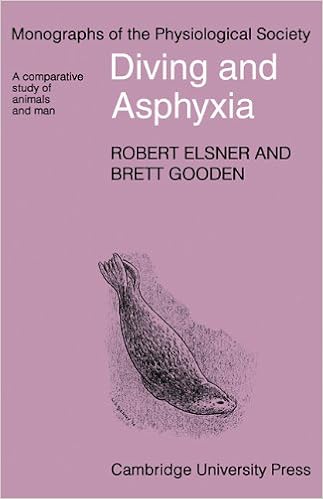
First revealed in 1983, this booklet matters the comparative physiological variations of vertebrate animals, particularly mammals, to cessation of respiring. those variations have been initially pointed out in species dwelling in aquatic habitats. The argument is gifted that the ordinary divers show a well-developed and very easily studied instance of a extra basic defence opposed to asphyxia.
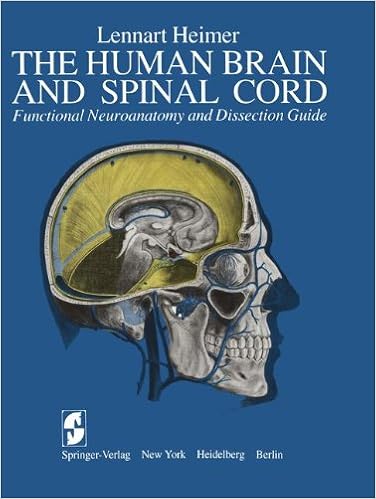
The Human Brain and Spinal Cord: Functional Neuroanatomy and Dissection Guide
This e-book used to be written to serve either as a consultant for the dissection of the human mind and as an illustrated compendium of the useful anatomy of the mind and spinal wire. during this feel, the e-book represents an up to date and multiplied model of the ebook The Human mind and Spinal twine written through the writer and released in Swedish via Scandinavian collage Books in 1961.
- Cell imaging techniques : methods and protocols
- Cellulase : types and action, mechanism, and uses
- Neurosteroid Effects in the Central Nervous System: The Role of the GABA-A Receptor (Frontiers in Neuroscience)
- Molecular Cytogenetics. Protocols and Applications
- Attitudes on altitude: pioneers of medical research in Colorado's high mountains
Additional resources for Color Atlas of Surgical Anatomy for Esophageal Cancer
Sample text
Sakamoto Fig. 15 This is a subsequent photograph of the same specimen. The loose connective tissue is being cleared away by use of forceps. Fig. 16 Now the retropharyngeal space can be easily viewed. Thick septa connecting the prevertebral fascia and the visceral fascia, via the carotid sheath, are clearly visible. Note that these septa serve as a boundary for the retropharyngeal space. Fig. 15. Gentle clearing of the retropharyngeaI space (specimen 4) Fig. 16. Thick septa encircling the retropharyngeaI space (specimen 4) Illustrations and Photographs 15 16 43 44 T.
In the future, it is important to obtain a more definite and preoperatively applicable classification of tumor appearance that relates to the grade of malignancy. Strategy for Radical Lymph Node Dissection Huang [19] has stated that major improvement in survival has been achieved by detection OIf this disease in its early stages, and Ellis et al. " The importance of early diagnosis cannot be overemphasized. If the tumor is found as a carcinoma in situ, treatment is obviously much simpler. However, the majority of patients are still diagnosed in the advanced stage.
Sakamoto Right Deep Pathway Fig. 2 This is an illustration of the esophagus and related structures when approached from the right side, based on the dissection of specimen 1. As the arch of the azygos vein has been cut and moved away, the whole length of the thoracic esophagus can be seen. The lung has been shifted to the front. Thus, the back surface of the right bronchus as well as the inferior pulmonary vein are visible. The vagus nerve, after giving off the recurrent laryngeal nerve which surrounds the subclavian artery, descends along the trachea slightly obliquely and reaches the back side of the bronchus.