By Giampiero Ausili Cèfaro, Visit Amazon's Domenico Genovesi Page, search results, Learn about Author Central, Domenico Genovesi, , Carlos A. Perez
Defining organs in danger is an important job for radiation oncologists whilst aiming to optimize the advantage of radiation remedy, with supply of the utmost dose to the tumor quantity whereas sparing fit tissues. This e-book will turn out a useful advisor to the delineation of organs prone to toxicity in sufferers present process radiotherapy. the 1st and moment sections handle the anatomy of organs in danger, speak about the pathophysiology of radiation-induced harm, and current dose constraints and strategies for objective quantity delineation. The 3rd part is dedicated to the radiological anatomy of organs in danger as noticeable on common radiotherapy making plans CT scans, as a way to supporting the radiation oncologist to acknowledge and delineate those organs for every anatomical area – head and neck, mediastinum, stomach, and pelvis. The publication is meant either for younger radiation oncologists nonetheless in education and for his or her senior colleagues wishing to lessen intra-institutional adaptations in perform and thereby to standardize the definition of scientific objective volumes.
Read or Download Delineating Organs at Risk in Radiation Therapy PDF
Best anatomy books
Clinical Physiology and Pharmacology
This ebook is an obtainable number of case research situations perfect for body structure and pharmacology revision for pharmacy, scientific, biomedical technology, medical technology and healthcare scholars. truly based and arranged via significant organ approach, the booklet emphasises ways that key symptoms of ailment tell analysis and the alternative of therapy, including the suitable pharmacological mechanisms.
The Cytoskeleton, Vol. 1: Structure and Assembly
This quantity of the treatise offers with structural facets of the cytoskeleton: the features of the filaments and their parts; the association of the genes; motor proteins; interactions with membranes.
First revealed in 1983, this e-book issues the comparative physiological diversifications of vertebrate animals, specifically mammals, to cessation of respiring. those diversifications have been initially pointed out in species dwelling in aquatic habitats. The argument is gifted that the traditional divers exhibit a well-developed and with ease studied instance of a extra normal defence opposed to asphyxia.
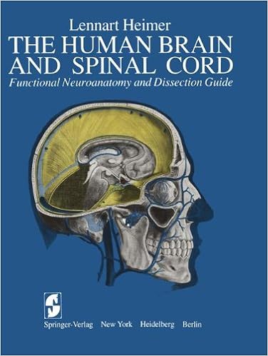
The Human Brain and Spinal Cord: Functional Neuroanatomy and Dissection Guide
This publication was once written to serve either as a consultant for the dissection of the human mind and as an illustrated compendium of the sensible anatomy of the mind and spinal twine. during this feel, the publication represents an up-to-date and extended model of the ebook The Human mind and Spinal twine written by means of the writer and released in Swedish via Scandinavian collage Books in 1961.
- Biology in Physics. Is Life Matter
- Essential Clinical Anatomy of the Nervous System
- Computational Strategies Towards Improved Protein Function Prophecy of Xylanases from Thermomyces lanuginosus
- Essential Clinical Anatomy of the Nervous System
Extra info for Delineating Organs at Risk in Radiation Therapy
Example text
Arch Intern Med 96:26–31 11. Goldgraber MD, Rubin CE, Palmer WL et al (1954) The early gastric response to irradiation; a serial biopsy study. Gastroenterology 27:1–20 12. Coia LR, Myerson RJ, Tepper JE (1995) Late effects of radiation therapy on the gastrointestinal tract. Int J Radiat Oncol Biol Phys 31:1213–1236 13. Balboni GC, Bastianini A, Brizzi E et al (1991) Anatomia Umana. Third Edition. Edi Ermes, Vol. 2, pp 116–144 14. Letschert JG, Lebesque J, Aleman JV et al (1994) The volume effect in radiation-related late small bowel complications: results of a clinical study of the EORTC Radiotherapy Cooperative Group in patients treated for rectal carcinoma.
The corpora cavernosa converge at the level of the subpubic arch, where their medial surfaces come into contact, with interposition of connective fascia (penile septum). The corpora cavernosa form a longitudinal groove both dorsally and ventrally, along the entire length of the penis corpus; the dorsal groove opens into the dorsal vein of the penis, whereas the ventral groove opens into the corpus spongiosum. Toward the distal terminations, the corpora cavernosa become thinner and terminate with a blunt apex encapsulated by the glans penis.
Actinic enteritis manifests as diarrhea and is variably associated with painful abdominal cramps, nausea, vomiting (rare), lack of appetite, and weight loss. Histological changes due to acute enteritis are particularly evident at the level of the mucosa. Apoptosis of cryptic cells occurs, determining a reduction in villi as well as of mucosa thickening, with subsequent inflammatory processes until crypt abscess formation. The damage, evident in the third week of radiotherapy, tends to recover spontaneously after 3–4 days from the end of radiotherapy course, to be completed within 12–14 days [15].


