By Dr. Peter S. Hechl, Dr. Reuben C. Setliff III, Prof. Dr. Manfred Tschabitscher (auth.)
For the newbie or for the finished sinus medical professional, getting to know the anatomy of the lateral nasal wall is an ongoing problem. even supposing there are very good usual anatomical references and both notable sinus classes with cadaver dissection, a reference depicting the surgical anatomy is required. A step by step surgical procedure at the anterior nasal backbone to the anterior wall of the sphenoid is gifted. The sinus health practitioner is faced with quite a lot of assorted areas created by way of the ethmoid bone. No different bone within the human physique has such a lot of anatomical diversifications. 4 serious anatomical constructions are emphasised because the starting place for an exact method of surgical procedure of the maxillary, anterior ethmoid, frontal, and posterior ethmoid sinuses. The objective of this e-book is to satisfy the great problem of supplying an anatomical technique with a view to serve the sinus health professional of each point of expertise and expertise.
Read Online or Download Endoscopic Anatomy of the Paranasal Sinuses PDF
Similar anatomy books
Clinical Physiology and Pharmacology
This booklet is an obtainable selection of case learn situations perfect for body structure and pharmacology revision for pharmacy, clinical, biomedical technological know-how, medical technology and healthcare scholars. in actual fact established and arranged via significant organ method, the publication emphasises ways that key signs of sickness tell prognosis and the alternative of therapy, including the suitable pharmacological mechanisms.
The Cytoskeleton, Vol. 1: Structure and Assembly
This quantity of the treatise bargains with structural elements of the cytoskeleton: the features of the filaments and their elements; the association of the genes; motor proteins; interactions with membranes.
First revealed in 1983, this e-book issues the comparative physiological diversifications of vertebrate animals, specifically mammals, to cessation of respiring. those variations have been initially pointed out in species residing in aquatic habitats. The argument is gifted that the ordinary divers exhibit a well-developed and comfortably studied instance of a extra normal defence opposed to asphyxia.
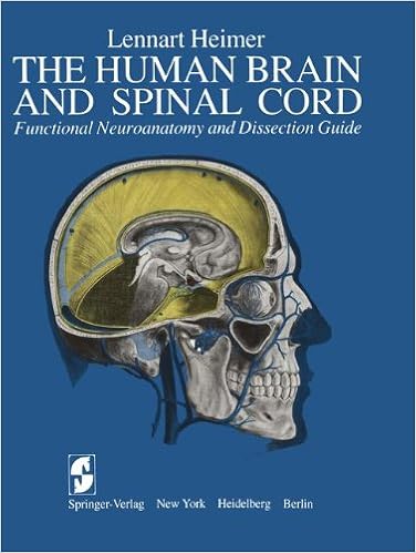
The Human Brain and Spinal Cord: Functional Neuroanatomy and Dissection Guide
This publication used to be written to serve either as a consultant for the dissection of the human mind and as an illustrated compendium of the useful anatomy of the mind and spinal twine. during this feel, the e-book represents an up to date and accelerated model of the publication The Human mind and Spinal wire written by way of the writer and released in Swedish by means of Scandinavian collage Books in 1961.
- The Human Nervous System
- An Illustrated Atlas of the Skeletal Muscles, 3rd Edition
- Laboratory Exercises in Anatomy and Physiology with Cat Dissections , Eighth Edition
- Pathology and Epidemiology of Cancer
- The Netter Collection of Medical Illustrations: Nervous System, Volume 7, Part II - Spinal Cord and Peripheral Motor and Sensory Systems, 2e
Extra info for Endoscopic Anatomy of the Paranasal Sinuses
Example text
Cut edge of uncinate process at 12 o'clock. ----------------------------~(]~---------------------------- 56 EO 6. The ethmoidal bulla Fig. 2. Right side, zero degree lens, ethmoidal bulla seen following partial resection of uncinate process. Fig. 3. Right side, zero degree lens, ethmoidal bulla; uncinate process has been resected; horizontal portion of basal lamella beneath bulla. --------------------------~[]~--------------------------- 6. The ethmoidal bulla EB 57 Fig. 4. Right side, 30 degree lens, uncinate process resected, ethmoidal bulla, and sinus lateralis.
The most consistent drainage pattern is from the posterior and medial aspect of the bulla medially into the hiatus semilunaris posterior, anterior to the basal lamella. The external anterior wall of the bulla serves as the posterior limit of the infundibulum. In many instances, a medial and inferior "teardrop" extension of the bulla may be present. The internal posterior anatomy is variable, sometimes presenting with a posterior bony wall and sometimes having the basal lamella as its posterior limit.
2. Left side, zero degree lens, drainage from most inferior and medial portion of middle meatus between middle turbinate and inferior turbinate. 1 1 \ \ \ SE MT , \ ,, ,, ,, , ... ;;1f \, ~\"'\ '" LNW ", , ,, NC Fig. 3. Right side, zero degree lens, middle meatus, a potential space only due to lateralization of middle turbinate. The middle meatus 31 MM Fig. 4. Right side, zero degree lens, middle meatus closed by nasal polyps. ,, ,, , I I MT ! \ II' \-\5 I 'I 1 I I ,, \ \ UP \ \ \ \ \ \ x Fig. 5.


