By Torsten Bert Moeller, Torsten Bert Möller, Emil Reif
Popular for its incredible illustrations and hugely functional details, the 3rd version of this vintage reference displays the very most modern in state of the art imaging expertise. including Volumes 1 and three, this compact and conveyable booklet offers a hugely really expert navigational device for clinicians looking to grasp the power to acknowledge anatomical buildings and safely interpret CT and MR images.Features:New CT and MR photos of the top qualityDidactic association utilizing two-page devices, with radiographs on one web page and full-color illustrations at the nextConcise, easy-to-read labeling on all figuresColor-coded, schematic diagrams that point out the extent of every sectionSectional enlargements for specified class of the anatomical structureComprehensive, compact, and conveyable, this publication is perfect to be used in either the school room and scientific surroundings.
Read Online or Download Pocket Atlas of Sectional Anatomy, Computed Tomography and Magnetic Resonance Imaging, Vol. 2: Thorax, Heart, Abdomen, and Pelvis PDF
Similar anatomy books
Clinical Physiology and Pharmacology
This ebook is an available selection of case learn eventualities perfect for body structure and pharmacology revision for pharmacy, clinical, biomedical technological know-how, scientific technology and healthcare scholars. truly dependent and arranged through significant organ procedure, the e-book emphasises ways that key signs of ailment tell analysis and the alternative of remedy, including the proper pharmacological mechanisms.
The Cytoskeleton, Vol. 1: Structure and Assembly
This quantity of the treatise bargains with structural elements of the cytoskeleton: the features of the filaments and their parts; the association of the genes; motor proteins; interactions with membranes.
First revealed in 1983, this booklet matters the comparative physiological variations of vertebrate animals, specially mammals, to cessation of respiring. those diversifications have been initially pointed out in species dwelling in aquatic habitats. The argument is gifted that the usual divers reveal a well-developed and comfortably studied instance of a extra normal defence opposed to asphyxia.
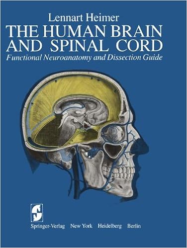
The Human Brain and Spinal Cord: Functional Neuroanatomy and Dissection Guide
This publication was once written to serve either as a consultant for the dissection of the human mind and as an illustrated compendium of the sensible anatomy of the mind and spinal twine. during this experience, the publication represents an up-to-date and multiplied model of the e-book The Human mind and Spinal wire written through the writer and released in Swedish via Scandinavian collage Books in 1961.
- Protein turnover and lysosome function
- Gray’s Anatomy Review
- The Fasciae: Anatomy, Dysfunction and Treatment
- Martin: Human Anatomy and Physiology
- High-yield gross anatomy
Additional resources for Pocket Atlas of Sectional Anatomy, Computed Tomography and Magnetic Resonance Imaging, Vol. 2: Thorax, Heart, Abdomen, and Pelvis
Sample text
24. 25. 26. 37 Transverse sinus of pericardium Aortic bulb and aortic valve Right coronary artery Right atrium Right ventricle Right atrioventricular (tricuspid) valve Xyphoid process of sternum Right coronary artery (posterior interventricular branch and right marginal branch) Diaphragm Liver Esophagus Supraspinous ligament Interspinal ligaments Spinous process Spinal cord 27. 28. 29. 30. 31. 32. 33. 34. 35. 36. 37. 38. 39. 40. 41. 42. 43. 44. Bifurcation of trachea Right pulmonary artery Ligamenta flava Thoracic vertebra Left atrium Intervertebral space Coronary sinus Descending aorta Diaphragm (lumbar part) Paratracheal lymph nodes Juxtaesophageal lymph nodes Tracheobronchial lymph nodes Anterior mediastinal lymph nodes Prevertebral lymph nodes Parasternal lymph nodes Subpericardial adipose tissue Superior phrenic lymph nodes Inferior phrenic lymph nodes Moeller, Sectional Anatomy © 2007 Thieme All rights reserved.
21. 22. 23. 24. 25. 26. 27. 34 35 36 37 38 39 Pectoralis major muscle Right auricle Right coronary artery Right ventricle Right atrioventricular (tricuspid) valve Right coronary artery (posterior interventricular branch) Right coronary artery (terminal branch) Diaphragm Rectus abdominis muscle Liver Splenius cervicis and capitis muscle Semispinalis capitis muscle Longus colli muscle Erector spinae muscle Trapezius muscle Semispinalis thoracis muscle (multifidus muscle) 28. 29. 30. 31. 32. 33. 34.
Deep cervical lymph nodes 35. Superficial cervical lymph nodes 36. Supraclavicular lymph nodes 37. Pulmonal lymph nodes 38. Intercostal lymph nodes 39. Prepericardial lymph nodes Moeller, Sectional Anatomy © 2007 Thieme All rights reserved. Usage subject to terms and conditions of license. 44 MRI of the Thorax 35 36 37 1/2 3 38 4 39 6 10 —— = Borders of lung segment —— = Pericardium Left Lung 1+2 Apicoposterior segment of upper lobe 3. Anterior segment of upper lobe 4. Superior lingular segment 6.


