By Massimo Gallucci MD, Silvia Capoccia MD, Alessia Catalucci MD (auth.)
The English variation encompasses a few adjustments from the 1st ItaHan variation, which require an evidence. to begin with, a few imag es, particularly a few 3D reconstructions, were converted in an effort to lead them to clearer. Secondly, in contract with the writer, we now have disowned one among our statements within the preface to the Italian version. particularly, we've additional a quick introductory textual content for every part, when it comes to clarification to the anatomical and physiological notes. this could make it more straightforward for the reader to appreciate and seek advice from this Atlas. those changes derive from our adventure with the former version and are supposed to be an development thereof confidently, there'll be extra variants to stick with, in order that we may perhaps additional increase our paintings and continue ourselves busy on lone a few evenings. eventually, the advancements during this variation are a reminder to the reader that one shouldn't ever buy the 1st version of a piece. UAquila, January 2006 The Authors Preface to the Italian variation i've been desiring to put up an atlas of neuroradiologic cranio-encephaHc anatomy for a minimum of the decade. basic anatomy has continuously been of significant and fascinating curiosity to me. through the years, whereas getting ready lectures for my scholars, i've got regularly loved lingering on anatomical info that at the present time are rendered with superb realism by means of regimen diagnostic ima ging.
Read Online or Download Radiographic Atlas of Skull and Brain Anatomy PDF
Similar anatomy books
Clinical Physiology and Pharmacology
This e-book is an available choice of case examine eventualities excellent for body structure and pharmacology revision for pharmacy, clinical, biomedical technology, medical technological know-how and healthcare scholars. sincerely based and arranged by means of significant organ process, the booklet emphasises ways that key symptoms of disorder tell prognosis and the alternative of therapy, including the suitable pharmacological mechanisms.
The Cytoskeleton, Vol. 1: Structure and Assembly
This quantity of the treatise offers with structural features of the cytoskeleton: the features of the filaments and their elements; the association of the genes; motor proteins; interactions with membranes.
First revealed in 1983, this booklet matters the comparative physiological diversifications of vertebrate animals, specially mammals, to cessation of respiring. those variations have been initially pointed out in species residing in aquatic habitats. The argument is gifted that the usual divers show a well-developed and with ease studied instance of a extra basic defence opposed to asphyxia.
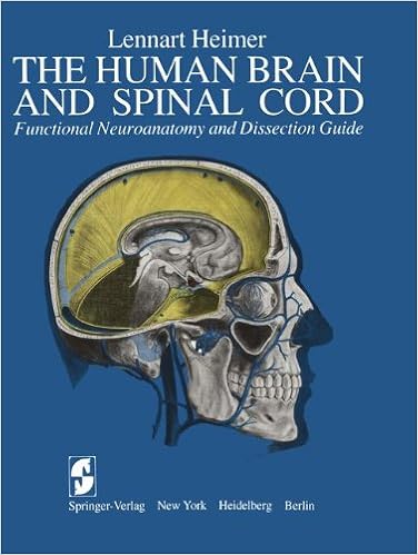
The Human Brain and Spinal Cord: Functional Neuroanatomy and Dissection Guide
This publication used to be written to serve either as a advisor for the dissection of the human mind and as an illustrated compendium of the sensible anatomy of the mind and spinal twine. during this experience, the e-book represents an up-to-date and improved model of the booklet The Human mind and Spinal wire written by means of the writer and released in Swedish by means of Scandinavian collage Books in 1961.
- Immunopharmacogenomics
- Biology of Sensory Systems, Second Edition
- Fundamental Anatomy for Operative General Surgery
- Anatomy Questions for the MRCS
Extra info for Radiographic Atlas of Skull and Brain Anatomy
Sample text
Below it, projection fibers arising from the cortex and directed towards the internal capsule, together with fibers ascending from below towards the cortex, form the "corona radiata". The former occupy a narrow space on each side of the bodies of the lateral ventricles. Similarly, a special contingent of fibers comes from the lateral geniculate body and reaches the occipital cortex, passing laterally to the occipital horn of the lateral ventricles, be- ing more horizontally oriented. e. the final part of the visual pathway.
Superior frontal sulcus CIngulate gyrus > Middle frontal gyrus (F2) Body of fornix ^ Superior precentral sulcus Body of lateral ventricle Precentral gyrus Circular insular sulcus »• Central or rolandic operculum Third ventricle Lateral fissure of Sylvius Short insular gyrus Superior temporal gyrus (Tl) Long insular gyrus Superior temporal sulcus Hippocampus Middle temporal gyrus (T2) Inferior temporal sulcus Inferior temporal gyrus (T3) Vertebral arteries - * Lateral occipitotemporal sulcus Temporal horn of lateral ventricle Parahippocampal gyrus (T5) Collateral sulcus (medial temporooccipital incisure) Fusiform gyrus (T4) Af^t4i ,^-_ ^ ^ 53 54 2 Sectional Anatomy of the Telencephalon i Superior sagittal sinus Longitudinal fissure / Superior precentral sulcus Superior frontal gyrus (F1) Precentral gyrus Cingulate sulcus Central sulcus Cingulate gyrus Postcentral gyrus Body of lateral ventricle Corona radiata Corpus callosum, isthmus Central or rolandic operculum Posterior transverse temporal gyrus Lateral fissure of Sylvius Superior temporal gyrus (T1) ' * Circular insular sulcus Anterior transverse temporal gyrus Short insular gyrus Superior temporal sulcus Long insular gyrus Middle temporal / gyrus n'2) Third ventricle Inferior temporal / sulcus Head of hippocampus Inferior temporal / gyrus (T3) ^ 4^ Lateral temporo-occipital sulcus Fusiform gyrus (T4) Parahippocampal gyrus (T5) #-r ^ B Coronal Sections Internal cerebral vein Cingulate gyrus Corpus callosum, isthmus Superior frontal gyrus (Fl) -^ Precentral gyrus Central gyrus Body of lateral j J ; ' / ventricle ^ ^ ^ Postcentral gyrus ^--^ Intraparietal sulcus Cingulate sulcus Fornix Supramarginal gyrus Lateral fissure of Sylvius .
Gyrus (T1) . * ! L-ji«pi3^i»** Parahippocampal gyrus (T5) /f, "^p^^ Head of hippocampus Fusiform gyrus (T4) Posterior transverse collateral sulcus ^ f V A Sagittal Sections -


